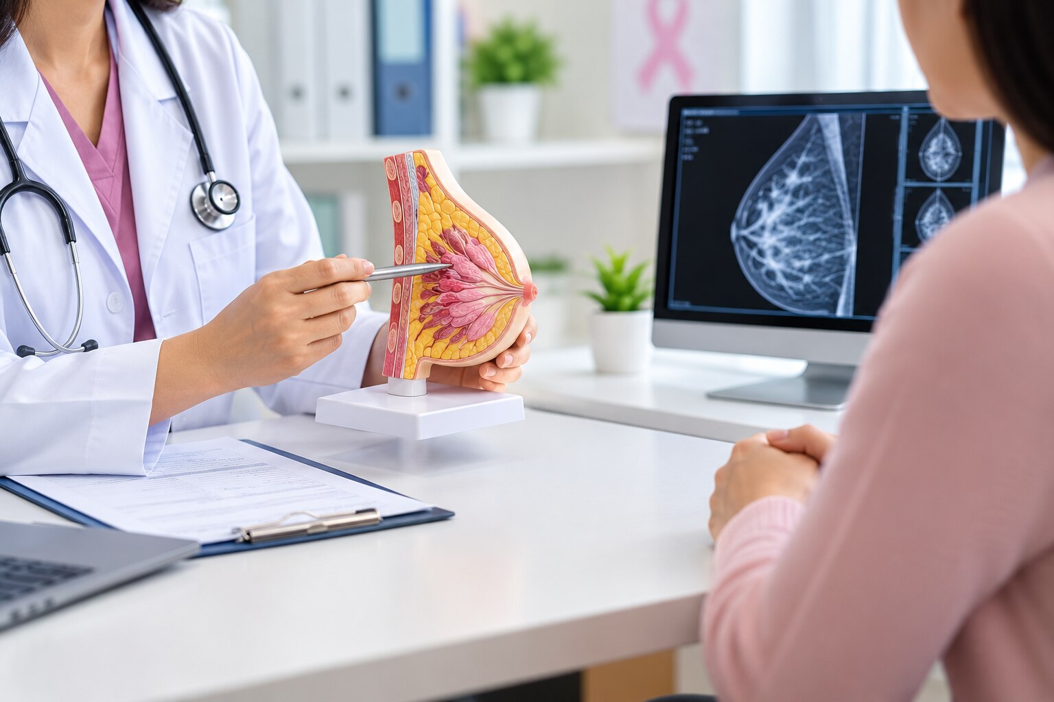
A breast check starts with your symptoms, medical history and a hands-on clinical examination—not automatically with a mammogram. You will learn what happens at the appointment, how to prepare, which test may follow, and when a reassuring examination still needs further review.
Key takeaways
- Expect questions about symptoms, history, medicines and breast-cancer risk.
- Tell the doctor when a change started and whether it varies with your cycle.
- Mammography, ultrasound or MRI depends on your age, symptoms and risk.
- Seek urgent assessment for a new lump, bloody discharge or skin changes.
What happens during a breast examination appointment?
A breast examination in Mumbai usually starts with questions, not a mammogram. The doctor reviews your symptoms, medical history and breast-cancer risk before examining you.
- You describe when a lump, pain or other change began and whether it is growing. Tell the doctor about nipple discharge, skin or nipple changes, recent injury, pregnancy or breastfeeding, menstrual timing, previous breast surgery or breast problems, and personal or family history of breast or ovarian cancer.
- The doctor explains the examination, answers questions and asks for your consent. You remove clothing from the waist up in private and use a gown or sheet; fasting is not needed for a hands-on clinical examination.
- While you sit or stand, the doctor inspects both breasts with your arms relaxed, raised, pressed against your hips and sometimes leaning forward. They look for differences in size or shape, swelling, skin dimpling, redness, visible veins, nipple inversion or other changes.
- You then lie down, usually with one arm above your head. Using the flat pads of their fingers, the doctor feels the breast tissue in a systematic pattern, checking for lumps, tenderness, firmness and mobility. They examine both breasts, the armpits and nearby chest area, and may gently check the nipples for discharge.
The appointment ends with an explanation of what was found. If a lump or symptom needs clarification, ask which imaging or follow-up is appropriate and when you will receive the result; a normal-feeling examination alone cannot rule out every breast problem.
What should you tell the doctor, and how should you prepare?
For a women breast health consultation in Mumbai, describe the change in your own words rather than waiting for the doctor to find it. State when a lump began, whether it is growing, and any breast or nipple pain, discharge, skin dimpling, redness, nipple inversion, rash or recent injury.
Mention pregnancy, breastfeeding, menstrual-cycle timing and hormone medicines.
Share these risk details clearly:
- Personal history of breast, ovarian or other related cancer
- Breast surgery, biopsy, cysts or previous abnormal imaging
- A mother, sister, daughter or other close relative with breast or ovarian cancer
- Several affected relatives, diagnosis at a young age, male breast cancer, cancer in both breasts, or a known familial pathogenic variant
A strong family pattern can lead to genetic counselling and enhanced surveillance instead of routine mammography alone.
Bring the following:
| Item | What to bring | Why it matters |
|---|---|---|
| Reports | Mammograms, ultrasounds, MRI or biopsy results | Allows comparison with earlier findings |
| History | Surgery and treatment records | Prevents missing important details |
| Medicines | Current prescriptions and hormone products | Helps interpret symptoms and risk |
| Identification | Registration or insurance documents | Supports appointment and referral records |
You do not need to fast for a hands-on examination. Wear a two-piece outfit for easier changing, and avoid deodorant or talcum powder if imaging is planned because residue can obscure mammogram findings. Ask who interprets imaging and how abnormal results or biopsies will be followed up.
Which breast test is right after the examination?
The right next test depends on what the clinician finds, your age, symptoms, pregnancy status and risk. A clinical breast examination in Mumbai is a hands-on assessment; it cannot rule out a small or deep cancer. Self-awareness helps you notice change, but it does not replace screening or examination.
| Option | What it means | When it applies |
|---|---|---|
| Breast self-awareness | Knowing your usual appearance and feel, then reporting a persistent or changing lump, discharge, pain or skin change | Every day; do not rely on formal monthly checks to reduce cancer risk |
| Clinical examination | Inspection and palpation of both breasts, armpits and nearby chest tissue | The starting assessment for a new concern |
| Mammography or tomosynthesis | X-ray imaging that can reveal masses or calcifications | Screening without symptoms, or diagnostic assessment; from age 40, usually with targeted ultrasound when needed |
| Ultrasound | Sound-wave imaging that distinguishes fluid-filled from solid areas | Usually first for a palpable mass under 30; also common during pregnancy or breastfeeding and alongside mammography at 30–39 |
| MRI | Contrast-enhanced imaging that shows blood flow and disease extent | Selected high-risk screening, staging or unresolved findings; not a routine replacement for mammography |
| Biopsy | Tissue sampling: core needle is commonly preferred for a solid lesion; fine-needle aspiration and surgical biopsy answer different questions | A suspicious or examination-imaging-discordant finding needs tissue diagnosis |
Pregnancy or breastfeeding does not make a concerning lump safe to ignore. Ultrasound is usually first, while mammography can follow when clinically indicated with radiation precautions; persistent or suspicious findings may require biopsy. A benign aspiration cannot dismiss a discordant result.
When should you seek urgent assessment, and who should coordinate it?
Do not wait for a routine screening appointment if you notice a new lump, bloody or clear discharge from one breast, new nipple inversion, skin dimpling, peau d’orange, persistent eczema-like nipple change, or a lump in the armpit. Persistent focal pain, especially with a lump, also needs prompt assessment.
| Change noticed | Action | Who should coordinate |
|---|---|---|
| New lump in the breast or armpit | Arrange prompt clinical assessment and appropriate imaging | Gynaecologist, breast surgeon, or breast clinic |
| Bloody or clear one-sided discharge | Seek prompt assessment rather than monitoring it at home | Clinician who can arrange imaging and referral |
| New inversion, dimpling, peau d’orange, or persistent nipple rash | Book an early appointment | Doctor with access to breast imaging and biopsy pathways |
| Persistent focal pain or a lump during breastfeeding | Arrange assessment; do not assume it is only hormonal or infectious | Clinician familiar with lactation and breast conditions |
When choosing care in Mumbai, check these points:
- Confirm the doctor’s registration and relevant training. “Breast examination doctor in mumbai” is a search phrase, not a recognised qualification.
- Ask who performs and interprets mammography, ultrasound, or MRI, and whether the radiologist regularly handles breast imaging.
- Ask how an abnormal result, recommended biopsy, or mismatch between symptoms and imaging will be followed up.
- A breast checkup gynecologist in mumbai can coordinate initial assessment and referral. Her Space by Dr. Shifa can be one starting point if the clinician provides a clear imaging and referral plan.
- Tell the team about implants, prior surgery, dense breasts, pregnancy, or breastfeeding before imaging.
Related service
GYNAECOLOGY by Dr. Shifa Anwar Panwala Cervical cancer screening is a vital preventive service that helps detect early changes in the cervix before they develop... View service → |
Why can a normal examination still require follow-up?
A normal-feeling examination does not prove that every part of the breast is normal. A small or deep lesion can escape palpation, and a lump that feels benign can still need imaging or tissue sampling when it persists, changes, or conflicts with your risk profile.
Arrange further assessment if any of these apply:
- A lump, focal pain, nipple discharge, skin change, or nipple retraction remains after the examination.
- You have a close relative with breast or ovarian cancer, several affected relatives, cancer diagnosed young, male breast cancer, bilateral disease, or a known inherited pathogenic variant.
- You have dense breast tissue, implants, previous breast surgery, or are breastfeeding, making examination or image interpretation harder.
- The examination identifies a concern but the mammogram or ultrasound is reported as normal, or the imaging finding does not explain what you or the clinician can feel.
A women breast health consultation in Mumbai should record inherited risk and decide whether you need genetic counselling, targeted imaging, enhanced surveillance, or biopsy rather than routine mammography alone.
Ask a breast examination doctor in Mumbai who performs and interprets the imaging, how discordant results will be reviewed, and who will follow up an abnormal scan or biopsy. Do not close the case because one test appears reassuring; persistent clinical concern needs reconciliation between the examination, radiologist and next investigation.
Frequently asked questions
What happens during a breast examination appointment?
The doctor asks about your symptoms, menstrual and pregnancy history, medicines, previous breast problems and family history. They inspect and feel both breasts and the lymph nodes near your armpits and collarbones.
What should you tell the doctor before a breast examination?
Describe the change, its location, when you noticed it, whether it is painful, and whether it changes with your menstrual cycle. Mention nipple discharge, prior breast surgery, pregnancies, hormone use and relatives with breast or ovarian cancer.
Which breast test is needed after the examination?
The doctor chooses between diagnostic mammography, breast ultrasound or breast MRI based on your age, examination findings, symptoms and breast-cancer risk. Imaging does not replace the clinical examination.
When should you seek urgent breast assessment?
Arrange prompt medical assessment for a new hard or persistent lump, bloody nipple discharge, nipple inversion, skin dimpling, an ulcer, or a red, hot, swollen breast with fever.
Why can a normal breast examination still need follow-up?
A clinical examination cannot detect every breast problem. Follow-up is needed when symptoms persist, imaging shows an abnormality, your risk is elevated, or the doctor asks you to return for a repeat assessment.
Keywords